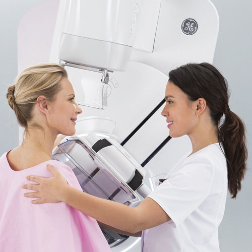
3D Screening Mammography
What is it and why should I consider it?
3D mammography is an important technological advancement that can improve early detection and improve your screening experience.
You already know that the mammogram is the most widely used and most trusted tool for breast screening. The goal of mammography is the early detection of breast cancer. When detected early, the chances for successfully treating breast cancer increase dramatically. In addition, finding cancer at an earlier stage may help save a woman’s breast by eliminating the need for a mastectomy.
3D mammography, or tomosynthesis, is a recent and significant innovation in breast imaging—approved by the FDA in 2011. Numerous European and American clinical studies have demonstrated that adding tomosynthesis to a screening mammogram increases the cancer detection rate by about 40% and significantly lowers recall rates.1 It has also been shown to find more invasive cancers earlier than traditional mammography2, which is why so many hospitals and imaging centers are adopting this technology.
3D Mammography at Desert Rose Women’s Center is:
- Safe and effective, with improved detection and fewer call-backs.
- Our GE Senographe Pristina offers the lowest radiation exposure of all 3D mammography systems (about the same exposure as a conventional 2D mammogram)
- Is faster than conventional mammograms with less patient discomfort
- Is performed with advanced computer-aided detection (CAD) technology
- Is less expensive than other providers, which can make a real difference if follow up testing is needed, or if you’re uninsured.
Are you due for your mammogram? Let us show you just how comfortable and easy it is to have a 3D mammogram at Desert Rose Women’s Center!
Current guidelines from the American Cancer Society (ACS), the American Medical Association (AMA) and the American College of Radiology (ACR) recommend screening mammography every year for women, beginning at age 40. The National Cancer Institute (NCI) recommends that women who have had breast cancer and those who are at increased risk due to a genetic history of breast cancer should seek expert medical advice about whether they should begin screening before age 40 and about the frequency of screening.
Before scheduling a mammogram, you should discuss problems in your breasts with your doctor. In addition, inform your doctor of hormone use, any prior surgeries, and family or personal history of breast cancer. Generally, the best time is one week following your period. Do not schedule your mammogram for the week before your period if your breasts are usually tender during this time. Always inform your x-ray technologist if there is any possibility that you are pregnant.
On the day of the exam:
- Do not wear lotion, deodorant, or powder under your arms or on your breasts.
- Describe any problems you’re experiencing with your breasts with your technologist.
- Remove all jewelry and clothing from the waist up. You will be given a gown that opens in the front.
To image your breast, a technologist will position you near the machine and your breast will be placed on a platform and compressed with a paddle. Breast compression is necessary* in order to:
- Spread out the tissue – so that small abnormalities won’t be obscured.
- Allow use of a lower x-ray dose.
- Even out the breast thickness – so that all of the tissue can be visualized.
- Hold the breast still – to eliminate blurring of the image caused by motion.
- Reduce x-ray scatter – to increase picture sharpness.
The technologist will go behind a glass shield while making the x-ray exposure. You will be asked to change positions slightly between views. The process is repeated for the other breast. Routine views are a top-to-bottom and side view.
*If you are having a 3D mammogram, less compression is typically required than a 2D mammogram.
A Screening Mammogram takes about 5 minutes; a Diagnostic Mammogram around half an hour. The technologist will apply compression on your breast and, as a result, you will feel pressure on the breast as it is squeezed by the compression paddle. Some women with sensitive breasts may experience some minor discomfort. Be sure to inform the technologist if pain occurs as compression is increased. If discomfort is significant, less compression will be used.
*If you are having a 3D mammogram, less compression is typically required than a 2D mammogram.
- Skaane P, Bandos AI, Gullien R, et al. Comparison of digital mammography alone and digital mammography plus tomosynthesis in a population-based screening program. Radiology. 2013;267(1):47-56
- Durand, Melissa A et al; Breast Cancer Screening Using Tomosynthesis in Combination with Digital Mammography. JAMA 2014.6095.jama/10.1001:doi. 2507-2499: (24)311; 2014

